
Dog Diagnostic Imaging
Diagnostic imaging is an important part of modern veterinary medicine and allows veterinarians to evaluate internal structures safely, efficiently, and non-invasively.
Dog Diagnostic Imaging Services
Many medical conditions cannot be diagnosed through a physical examination alone, which is why imaging technology often plays a critical role in identifying injuries, illness, pain, and chronic disease in dogs.
At Parkdale Animal Hospital, we use advanced diagnostic imaging tools to help assess a wide variety of medical conditions affecting dogs of all ages. Whether a dog is experiencing limping, breathing difficulties, vomiting, abdominal discomfort, or more subtle changes in behaviour or mobility, diagnostic imaging can provide valuable information that helps guide accurate diagnosis and treatment planning.
Our veterinary team understands that medical concerns involving internal disease can feel stressful and overwhelming for owners. We are committed to providing compassionate care, thorough diagnostics, and clear communication throughout the diagnostic process so families understand both the findings and the recommended next steps.
What Is Diagnostic Imaging?
Diagnostic imaging refers to technologies that allow veterinarians to visualize structures inside the body without surgery or invasive procedures. These tools help veterinarians assess bones, joints, organs, lungs, soft tissues, and other internal structures that cannot be fully evaluated externally.
Imaging is often used alongside physical examinations, laboratory testing, and medical history to create a more complete understanding of a patient’s condition.
At Parkdale Animal Hospital, our diagnostic imaging services may include digital radiography (X-rays) and ultrasound examinations. Each imaging type provides different types of information, and many patients benefit from a combination of diagnostic tools depending on their symptoms and medical concerns.
Diagnostic imaging is commonly used for both emergency conditions and ongoing chronic disease monitoring. In many cases, early imaging allows problems to be identified sooner, which may improve treatment options and long-term outcomes.
When Diagnostic Imaging May Be Recommended
Diagnostic imaging may be recommended whenever veterinarians need additional information about structures inside the body. Some conditions are obvious externally, while others require imaging to identify the underlying cause accurately.
Dogs experiencing limping, mobility changes, or suspected injuries may require imaging to evaluate bones, joints, and soft tissues. Imaging is also commonly used when dogs develop vomiting, diarrhea, abdominal pain, breathing difficulties, coughing, urinary symptoms, or unexplained behavioural changes.
Other situations where diagnostic imaging may be recommended include suspected foreign body ingestion, arthritis evaluation, bladder stones, cancer screening, pregnancy assessment, neurologic symptoms, or monitoring progression of chronic disease.
Sometimes imaging is recommended even when symptoms appear relatively mild because internal disease can occasionally progress silently before becoming more obvious clinically.
Our veterinary team carefully selects diagnostic recommendations based on each patient’s symptoms, examination findings, medical history, and overall health status.
Digital Radiography (X-Rays) for Dogs
Digital radiography, commonly called X-rays, is one of the most frequently used imaging tools in veterinary medicine.
X-rays use low levels of radiation to create detailed images of structures inside the body. Digital radiography provides rapid image acquisition and allows veterinarians to evaluate images immediately, improving efficiency and reducing wait times.
Radiographs are particularly valuable for evaluating bones and joints. Dogs with limping, suspected fractures, arthritis, spinal pain, or traumatic injuries often benefit from X-ray imaging to help identify abnormalities affecting the skeletal system.
Chest radiographs are commonly used to assess the lungs, airways, and heart. X-rays may help identify pneumonia, heart enlargement, fluid accumulation, certain tumors, or other respiratory conditions contributing to coughing or breathing difficulty.
Abdominal radiographs can help evaluate organ size and shape while also identifying conditions such as intestinal obstruction, swallowed foreign material, constipation, bladder stones, or abdominal masses.
Dental radiographs are another important component of veterinary imaging because many painful dental conditions occur beneath the gumline where they cannot be seen during a visual examination alone. Click here for more information about dental care at Parkdale Animal Hospital.
Digital imaging technology allows veterinarians to enhance and review images in detail while minimizing radiation exposure for both patients and staff.
Ultrasound Examinations in Dogs
Ultrasound is another valuable diagnostic imaging tool commonly used in veterinary medicine.
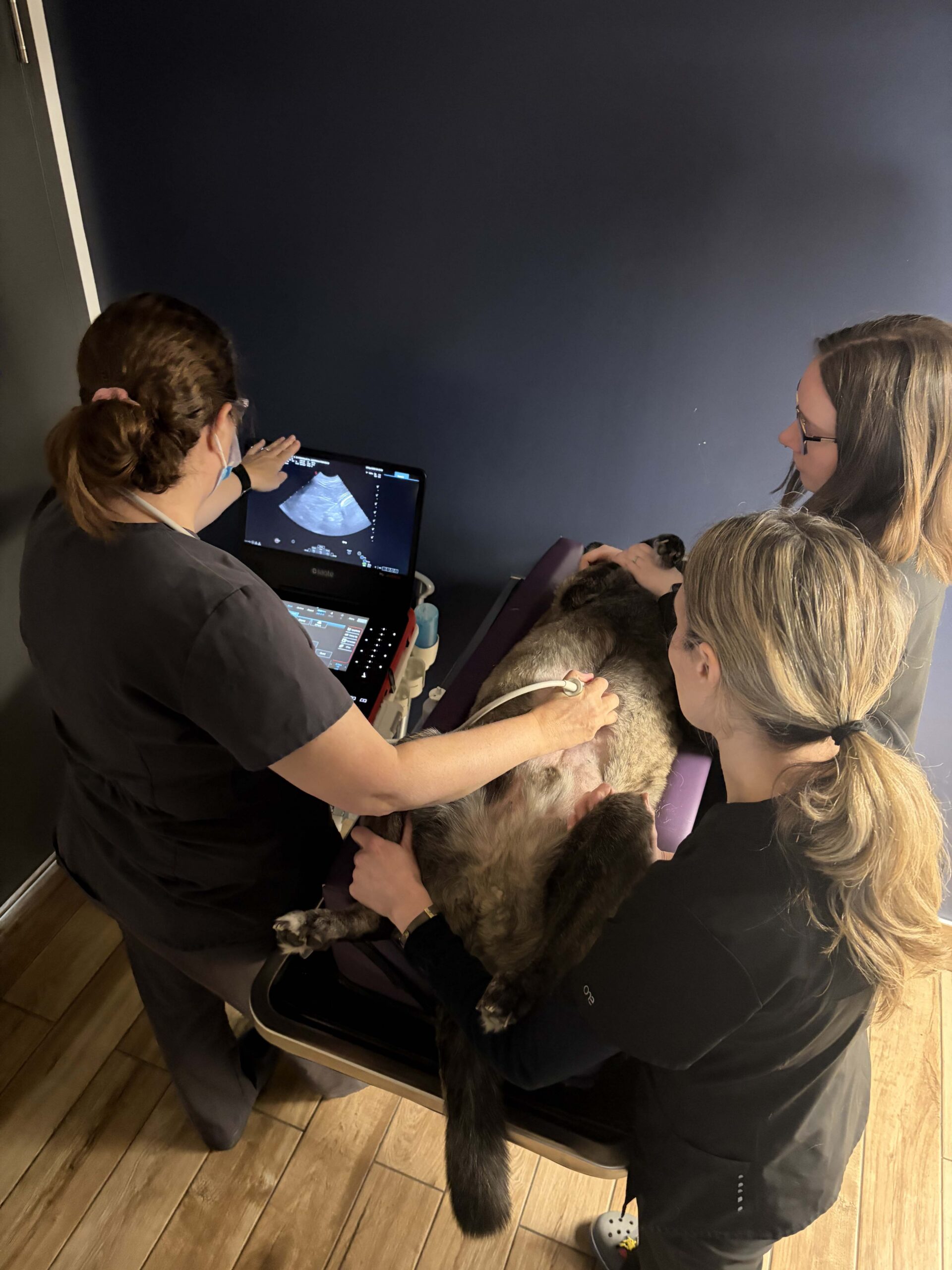
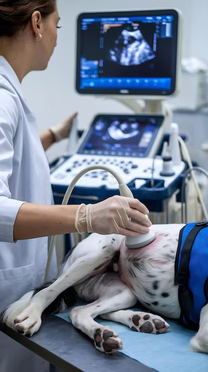
Unlike radiographs, which create static images, ultrasound uses sound waves to produce real-time images of soft tissues and internal organs. This allows veterinarians to evaluate organ structure, tissue texture, fluid accumulation, and movement within the body.
Ultrasound is especially useful for assessing abdominal organs such as the liver, kidneys, spleen, bladder, intestines, and pancreas. It may help identify inflammation, masses, cysts, stones, abnormal fluid, or structural abnormalities that are difficult to evaluate fully with radiographs alone.
Ultrasound examinations are also commonly used during pregnancy evaluations and may assist with monitoring fetal development.
In some cases, ultrasound can help guide diagnostic procedures such as fine needle aspirates or fluid collection by allowing veterinarians to visualize the target area in real time.
Because ultrasound evaluates soft tissues differently than X-rays, the two imaging methods are often complementary rather than interchangeable.
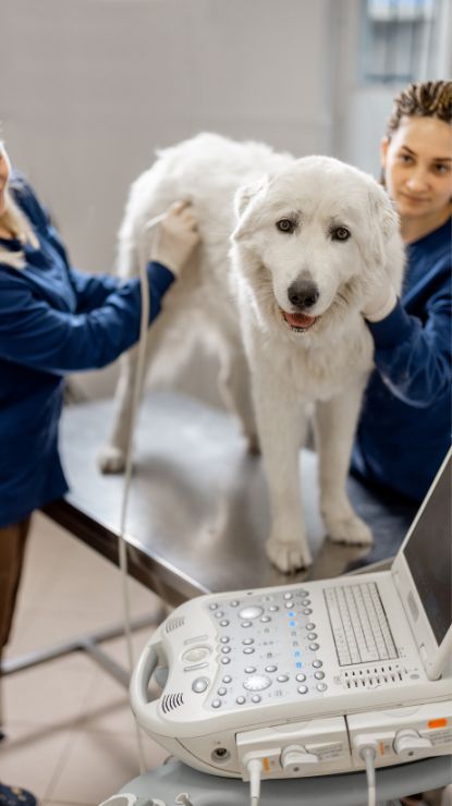
Some dogs tolerate ultrasound examinations very well while awake, while others may require mild sedation to reduce stress and improve image quality. Patient comfort and safety are always important considerations during the procedure.
Diagnostic Imaging for Lameness and Orthopedic Problems
Limping and mobility concerns are among the most common reasons dogs undergo diagnostic imaging.
Dogs may develop lameness due to arthritis, ligament injuries, fractures, hip dysplasia, spinal disease, joint instability, or soft tissue injuries. Sometimes symptoms develop suddenly after trauma, while other conditions progress gradually over time.
Radiographs allow veterinarians to evaluate bones, joints, and spinal alignment while looking for signs of degenerative disease, injury, or structural abnormalities.
In some cases, imaging may identify chronic changes that owners were unaware had been developing slowly over months or years.
Early diagnosis of orthopedic disease often allows for more effective pain management and long-term treatment planning aimed at preserving comfort and mobility.
Imaging for Gastrointestinal and Abdominal Disease
Diagnostic imaging is frequently used to investigate vomiting, diarrhea, abdominal pain, appetite changes, or unexplained weight loss.
Abdominal radiographs may help identify intestinal obstruction, foreign body ingestion, constipation, organ enlargement, or bladder stones. Ultrasound can provide more detailed information about organ structure and intestinal health while helping assess conditions such as pancreatitis, gastrointestinal inflammation, liver disease, or abdominal masses.
Foreign body ingestion is a particularly important concern in dogs because many dogs will swallow inappropriate objects such as toys, socks, bones, rocks, or fabric materials. Imaging often plays a critical role in determining whether surgery or other intervention may be necessary.
Because gastrointestinal symptoms can result from many different underlying conditions, imaging is often combined with bloodwork, fecal testing, and physical examination findings to reach an accurate diagnosis.
Diagnostic Imaging in Emergency Situations
Diagnostic imaging is often essential during veterinary emergencies.
Rapid imaging may help identify internal bleeding, traumatic injuries, foreign body obstruction, severe respiratory disease, or other life-threatening conditions requiring immediate treatment.
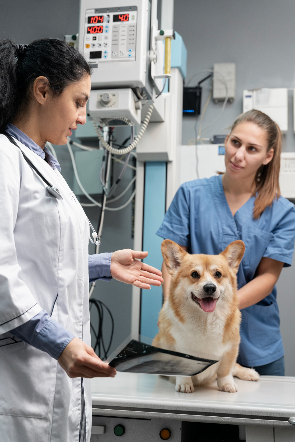
Dogs involved in accidents or traumatic injuries may require radiographs to assess fractures, lung injury, or internal organ damage. Patients experiencing difficulty breathing may need chest imaging to evaluate the lungs and heart quickly.
In emergency situations, rapid diagnosis can significantly influence treatment decisions and patient outcomes.
Our veterinary team works efficiently to stabilize patients while obtaining the diagnostic information needed to guide appropriate care.
Imaging and Heart Disease Evaluation
Imaging also plays an important role in evaluating heart disease in dogs.
Chest radiographs may help assess heart size and identify fluid accumulation within the lungs associated with congestive heart failure. Ultrasound examinations of the heart, called echocardiograms, provide more detailed information about heart structure and function.
At Parkdale Animal Hospital, we collaborate with a traveling board-certified veterinary cardiologist who provides advanced cardiac consultation and echocardiography services at our hospital. This allows many dogs to receive specialized cardiac evaluation in a familiar environment without traveling to a referral center.
Advanced cardiac imaging can be especially valuable for dogs with heart murmurs, arrhythmias, coughing, exercise intolerance, or suspected cardiac disease.
Patient Comfort and Safety During Imaging
Patient comfort and safety are important priorities throughout all imaging procedures.
Many dogs tolerate diagnostic imaging extremely well with gentle handling and positioning. However, some patients experiencing pain, anxiety, or stress may benefit from mild sedation to improve comfort and allow for safer image acquisition.
Sedation may also improve image quality by reducing movement during the procedure, particularly for ultrasound examinations or orthopedic radiographs requiring precise positioning.
Our veterinary team carefully evaluates each patient individually and recommends sedation only when appropriate and beneficial.
Throughout every procedure, we strive to minimize stress while maintaining a calm and compassionate environment for both pets and owners.

Why Early Diagnosis Matters
Early detection often plays a major role in improving treatment options and long-term outcomes.
Many medical conditions become easier to manage when identified earlier in the disease process before severe complications develop. Diagnostic imaging helps veterinarians identify abnormalities sooner, allowing treatment plans to be developed more quickly and accurately.
Even chronic conditions such as arthritis, heart disease, or certain cancers may benefit from earlier diagnosis and ongoing monitoring.
In some situations, imaging can also provide reassurance by helping rule out serious disease when symptoms are mild or nonspecific.
Compassionate Diagnostic Care at Parkdale Animal Hospital
At Parkdale Animal Hospital, we are committed to providing thorough diagnostic evaluations combined with compassionate, individualized patient care. We understand that diagnostic testing can sometimes feel overwhelming, especially when pets are ill or uncomfortable.
Our veterinary team takes the time to explain imaging findings clearly, discuss treatment recommendations, and answer questions so owners feel informed and supported throughout the process.
Whether your dog requires imaging for an acute injury, chronic medical condition, or preventive evaluation, our goal is always to provide thoughtful, evidence-based care focused on improving health, comfort, and quality of life.
If your dog is experiencing pain, illness, mobility changes, breathing difficulty, gastrointestinal symptoms, or other concerning signs, contact our team today at 519-885-1200 or click here to schedule an examination.